 |
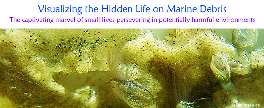
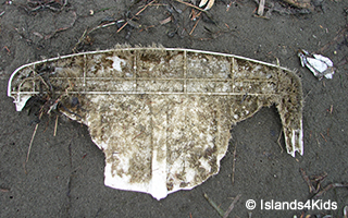
| The beautiful half-arches that appear to shoot out of the shells are predaceous organs of baby Gooseneck Barnacles (lepas anatifera). |
 |
 |
| |
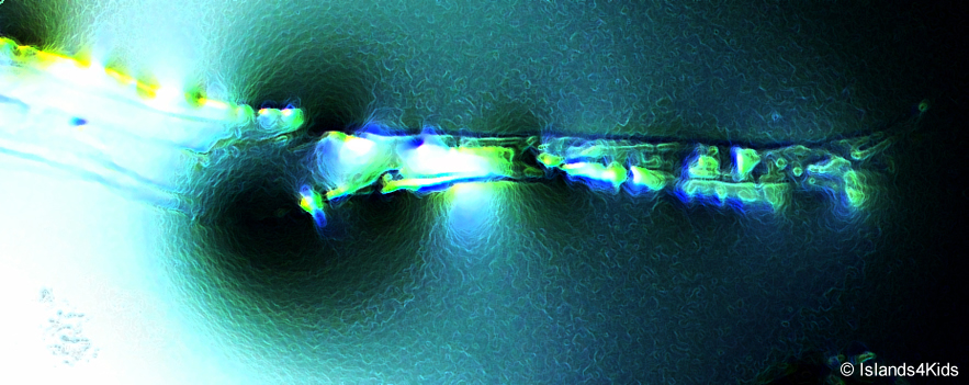 |
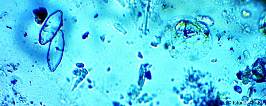
| This image is reminiscent of a sharp biotech sword gleaming on a pitch-black night. However, this is a microscopic image of Nitzschia peseudoseriata. The cell size is 8µm wide and 130-170µm in length. |
 |
|
| |
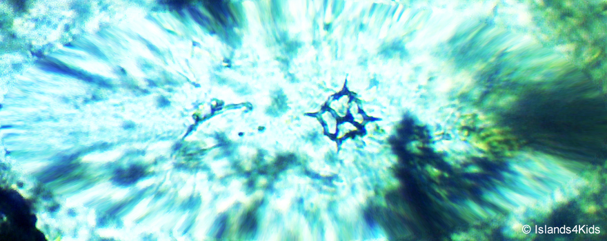 |
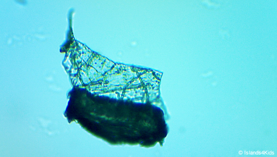
| Watch out! Someboday is throwing a ninja star! Actually, this is a Dictyocha / silicofkagellates (46µm) of phytoplankton. These are unbelievably cool and well-designed creatures. |
 |
|
| |
 |
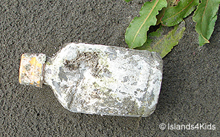
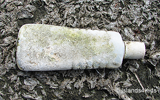
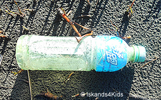
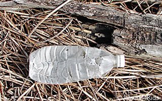
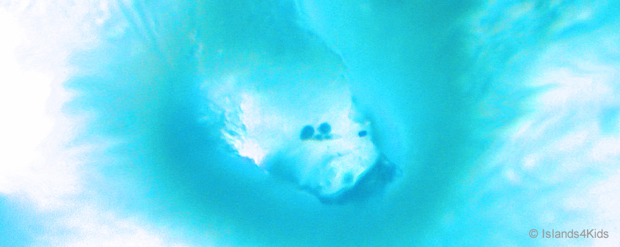
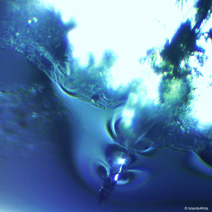
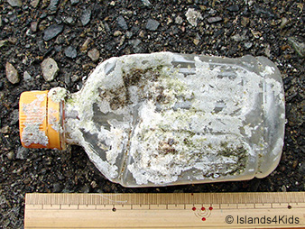
This photograph looks like a dramatic panoramic view of the ocean.
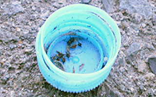
However, this photo is not a wide-angle shot of the ocean, but a close-up picture inside of a blue plastic bottle cap.
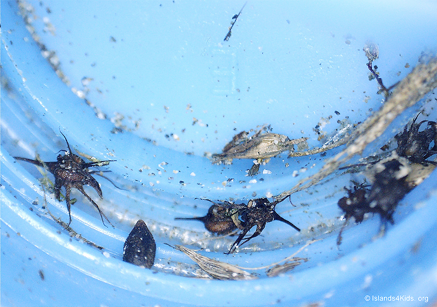
Nine unique zooplanktons once lived and traveled together in the Pacific Ocean on this 3cm diameter bottle cap until it washed ashore on the beach of Ocean Shores.
They were all dried at the time they were discovered, but their visible outlines (shapes and colors) were well preserved.
We have not yet determined the biological classification of these 3 creatures, but they seem to be a family (or order) of Branchyura Zoea (larva of crab) from their body and distinguishing branching legs.
Two small baby fish (about 2mm); one on top and the other on the bottom. They appear to be either Butterfish or Golden threadfin bream. |
 |
|
| |
 |
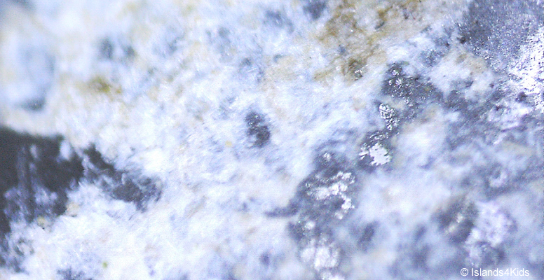
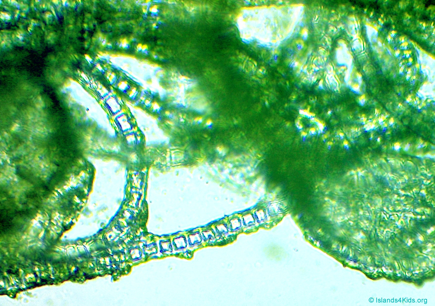
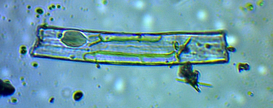
| The partition lines of each compartment are the protective outer coverings of Polyzoa. The median size of each compartment is 300µm x 800µm. This photo is taken with a polarization of kelly green filter set to deliver a clear image. |
 |
|
| |
 |
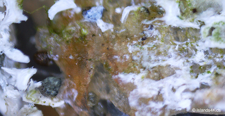
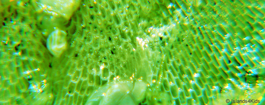
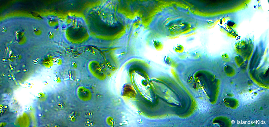
| What are you really?
This phytoplankton looks remarkably like an alien that has Zygomatic arch organs that provide CO2 to the entire body of the host as food. However, these are actually an alley of filamentous algae. Each cell diameter is about 45μm x 36μm. |
 |
|
| |
 |
| A miniture cosmic world projected on High Resolution Digital microscope screen. |
 |
|
| |
 |
| Two Astromphalus Algae (40µm) are enjoying a free ride on the head of a white ghost dragon swimming into votex rolls. |
 |
|
| |
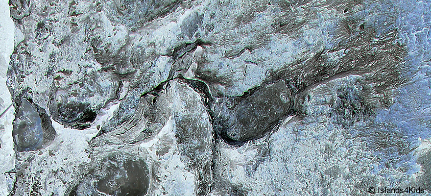 |
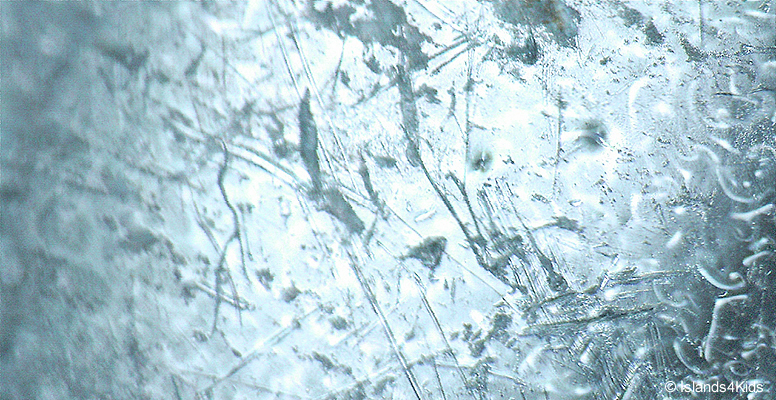
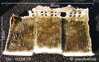
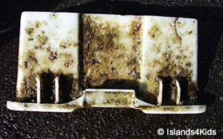
| Young oyster shells that were attached to the surface of a floating deck made of styrofoam had began developing colonies, which contain many nooks and crannies.
This magnified dynamic image looks like an expanse of glacier ice and glacial erosion. This floating deck may have been used at oyster cultivation farms in Japan. |
 |
|
| |
 |
| While preparing to observe Prorocentrum sp (Dinophyceae), we accidentally knocked the microscope, resulting in one of the Prorocentrum (size: 20µm x 60µm) on the glass to seemingly dive into the immersion oil. This gave us a surprisingly dynamic image, as shown above. |
 |
|
| |
 |
| What are you?
Over the course of microscope obsevations, we see many interesting images created accidentally by unknown means. |
 |
|
| |
 |
| A school of phytoplanktons. How many species of planktons can you find? |
 |
|
| |
 |
| Planctnema chlorophyceae |
|
|
| |
| |
| |
|
|
| |
| |
| |
|
|
| |
| |
| |
|
|
| |
| |
| |
|